
Anatomy of the abdomen and male pelvis using cross-sectional imaging - interactive atlas of human anatomy
Anatomy of the abdomen and male pelvis using cross-sectional imaging - interactive atlas of human anatomy
We created an anatomical atlas of abdominal and pelvic CT which is an interactive tool for studying the conventional anatomy of the normal structures based on a multidetector computed tomography. Anatomical structures of the abdomen and pelvis are visible as interactive labeled images.
Cross sectional anatomy: MDCT of the abdomen and pelvis
An enhanced (portal venous phase – 70 seconds) multidetector computed tomography was performed on a healthy subject in axial plane with coronal and sagittal reformatted images.
Data and DICOM images stocked on our PACS (Picture Archiving and Communicating System) were processed and exported as JPEG images. Adobe Animate and Adobe Photoshop allowed us to develop an atlas-based application with useful features and user-friendly interface for the study of the abdomen and pelvis anatomy (labeled according to the Terminologia Anatomica).
Anatomy of the abdominal cavity and the male pelvis: how to view anatomical labels
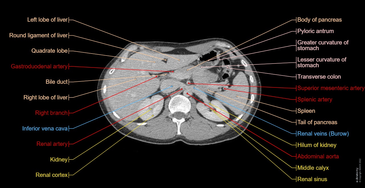
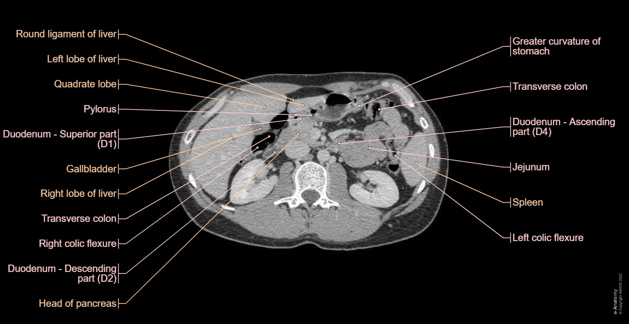
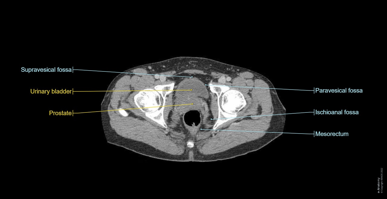
This tool provides access to a CT atlas in the axial plane, allowing the user to interactively learn abdominal anatomy. The images are labeled, providing an invaluable medical tool. The quiz mode provides evaluation of user progress.
On "Anatomical parts" the user can access the different groups of anatomical labels:
- Organs: liver, spleen, suprarenal gland, pancreas, gallbladder,
- Digestive tract: oesophagus, stomach, pyloric antrum, duodenum, jejunum, ileum, colon (caecum, ascending, transverse, descending colon, right and left colic flexure, sigmoid), rectum
- Urinary system: kidney (renal cortex and sinus), ureter, urinary bladder, prostate, urethra
- Arteries: abdominal aorta, iliac arteries, mesenteric arteries (inferior and superior), renal, splenic, heptatic, gastroduondenal, cystic arteries
- Veins: gonadic (testicular), iliac, epigastric, pudendal, gluteal, renal, hemiazygos veins, inferior vena cava
- Portal system: mesenteric (inferior and superior), splenic, hepatic portal veins
- Muscles, fascia, diaphragm
- Bones: ilium, sacrum, ribs
- Nerves
- Peritoneal cavity and spaces
- Lymph nodes regions
By moving the mouse cursor over a particular anatomical area of the liver, this liver segment is highlighted and labels are displayed: this feature was chosen to show hepatic segmentation:
- Segment 1: posterior segment – caudate lobe
- Segment 2: left posterior lateral segment
- Segment 3: left anterior lateral segment
- Segment 4: left medial segment
- Segment 5: anterior medial segment
- Segment 6: anterior lateral segment
- Segment 7: posterior lateral segment
- Segment 8: posterior medial segment
The vertical menu on the left shows coronal and sagittal reformatted images of the abdomen and pelvis, with digestive medical illustrations based on a three dimensional (3D) model (volume rendering of the CT)
This module is a comprehensive and affordable learning tool for radiology and medical students and residents and especially for radiologists, surgeons and gastrologists
There is no content here